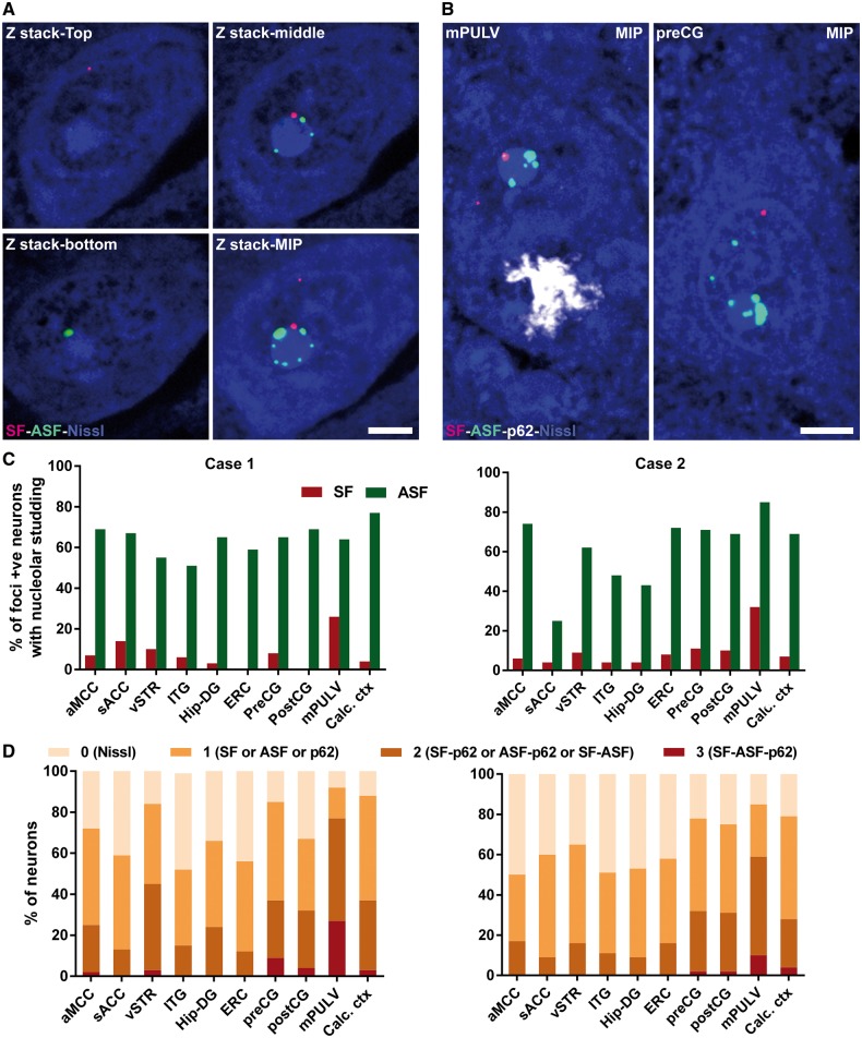
Figure 3.
Single neurons in degenerate brain regions harbour multiple C9orf72-associated inclusions. (A) Representative individual z-stack and maximum intensity projection (MIP) images from a single mPULV thalamic neuron show perinucleolar RNA foci. (B) MIP images from the mPULV and preCG show representative individual neurons with multiple types of inclusions. Scale bar = 5 µm for A and B. (C) Bar graphs depict the proportion of neurons in which at least one RNA focus directly abuts the nucleolus; this ‘nucleolar studding’ was more frequent for antisense than sense foci. (D) Stacked bars show the proportion of neurons harbouring 0, 1, 2, or 3 C9orf72-specific inclusion types across regions for Cases 1 and 2. In both patients, mPULV stood out as having the fewest neurons with no inclusions and the most with 2 or 3 inclusions. Direct statistical comparison between mPULV and non-PULV regions showed higher pathological multiplicity in mPULV (P < 0.001, see ‘Results’ section). aMCC = anterior midcingulate cortex; preCG = precentral gyrus; postCG = postcentral gyrus; ITG = inferior temporal gyrus; ERC = entorhinal cortex; vSTR = ventral striatum; sACC = subgenual anterior cingulate cortex; Calc ctx = calcarine cortex; Hip-DG = dentate gyrus of hippocampus.

