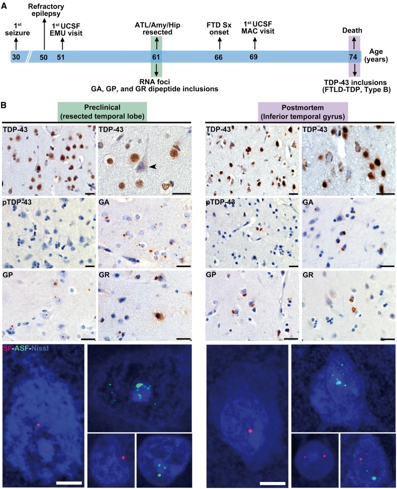
Figure 4.
C9orf72-specific pathological inclusions precede TDP-43 aggregation. (A) Schematic shows clinical milestones (above) and microscopic neuropathological features (below) in reference to the patient’s age timeline. (B) The resected left anterior temporal lobe represents the preclinical stage and is compared to post-mortem tissue from the contralateral anterior inferior temporal lobe. In the resected left temporal lobe, TDP-43 immunostaining revealed only a single NCI and no dystrophic neurites (Fig. 5). Intriguingly, numerous neurons lacking normal nuclear TDP-43 were observed in the apparent absence of a TDP-43 inclusion (arrowhead). These findings stand in contrast to the well-developed FTLD-TDP, type B, seen at autopsy. RAN-translated GA, GP and GR inclusions, as well as sense and antisense RNA foci, were conspicuous in both surgically resected tissue and post-mortem brain. Images with RNA foci are maximum intensity projections of a z-stack image to show the approximate abundance of foci within individual nuclei. Scale bars = 25 µm for bright field and 5 µm for fluorescent images. FTLD = frontotemporal lobar degeneration; DN = dystrophic neurites; RAN = repeat associated non-ATG; MAC = Memory and Aging Center; UCSF = University of California, San Francisco; ATL = anterior temporal lobe; EMU = Epilepsy Monitoring Unit.

