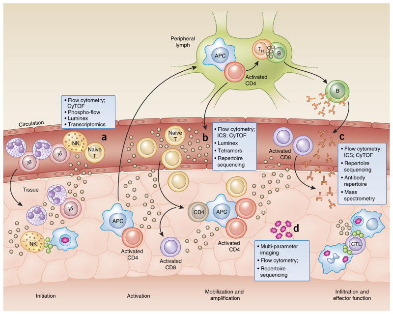
Figure 1.
Tools of the trade: a systems-immunology view. Investigating the immune system can be accomplished with a variety of techniques applied to the analysis of peripheral blood samples obtained from healthy donors, as well as those obtained at specific time points during infection, disease or after vaccination. Innate cell subsets can be characterized by RNA-seq, flow cytometry, cytometry by time-of-flight (CyTOF) and flow cytometry of phosphorylated proteins (Phospho-flow) to reveal mediators of early inflammation, and their signaling pathways (a). Cells can be classified by phenotype and functional capacity on the basis of a variety of cell-surface markers and receptor expression. Effector proteins, such as cytokines and cytolytic granules, along with other products of activated cells, can be detected intracellularly by flow cytometry or in the serum with other methodologies, such as bead-based multiplex assays (Luminex) or immunoassay kits (from Mesoscale Discovery), to rapidly survey more than 100 analytes in a single, small sample (b). Cell subsets of the adaptive immune system can be investigated for antigen specificity by tetramer technology and mass spectrometry to explore peptide presentation, as well as sequencing of the TCR and BCR repertoire for the identification of clonally expanded populations. Furthermore, antibody-repertoire sequencing can be used to characterize the extent of isotype class switching and affinity maturation (c). Many of these techniques are currently being adapted for the analysis of smaller tissue-biopsy samples to extract information about the local inflammatory environment and the infiltrating cell populations (d). s APC, antigen-presenting cell; TH, helper T cell; B, B cell; CD4, CD4+ T cell; NK, natural killer cell; γδ, γδ T cell; T, T cell; ICS, intracellular cytokine staining; CD8, CD8+ T cell; CTL, cytotoxic T lymphocyte.

