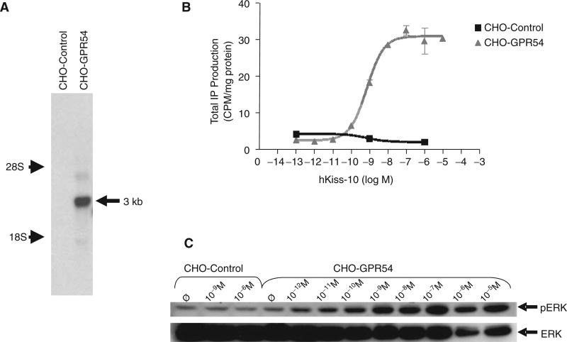
Fig. 1.
Characterization of pIRESneo3(hGPR54)- and pIRESneo3-transfected stable Chinese hamster ovary (CHO) cells (CHO-GPR54 and CHO-control, respectively). (A) Northern blot analysis of total RNA extracted from CHO-GPR54 and CHO-control cells, hybridized with a GPR54 riboprobe. A band was present in the CHO-GPR54 cell line but not in the CHO-control cell line. The band (indicated by the arrow) was estimated to be 3 kb in size based on the positions of 28S and 18S rRNA bands, consistent with the size of hGPR54 cDNA. (B) Stimulation of inositol phosphate (IP) production by increasing concentrations of hkiss-10 in the CHO-GPR54 cell line with an EC50 of 1.0 nM. CHO-control cells showed no response to hkiss-10. Symbols and error bars represent the mean ± SE of 3 replicates from a representative experiment. (C) Stimulation of ERK1/2 phosphorylation by increasing concentrations of kisspeptin in CHO-GPR54 and CHO-control cell lines. Cells were deprived of serum for 24 h, followed by stimulation with increasing concentrations (10−12 M to 10−5 M) of hkiss-10 for 5 min. Cells were lysed, extracts were isolated, and Western blot analysis was performed. Membranes were reprobed with ERK1/2 antibody to correct for protein loading. Samples were loaded in singlet; a representative blot is shown.

