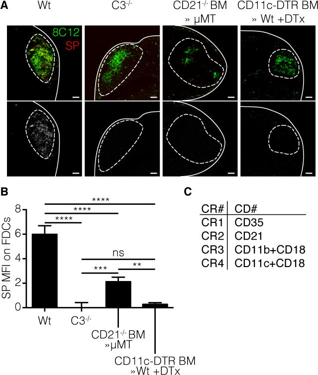
Figure 3. DCs and B cells collaborate in the transport of SP to the FDC.

Mice were injected with fluorescent labeled SP(red) in the footpad as described earlier. Four different mouse strains were used; wild type (Wt), complement C3 knockout (C3−/−), complement receptor 2 (CD21) knockout bone marrow (BM) in μMT B cell deficient recipients (CD21−/− BM≫μMT) and CD11c-dtr BM in Wt recipients (CD11c-dtr BM≫Wt) injected with Dtx 48h prior to immunization. The FDC network was stained using 8C12 (green). a | Confocal micrographs of pLN 12h after immunization with SP. Line indicates LN capsule, dotted line indicates follicle area. Bottom row: greyscale image of the SP channel. Scale bars 100 μm b | Quantification of confocal images by Cell Profiler software. Only co-localized signal was measured. **** p<0.0001, *** p<0.001, ** p<0.01, ns not significant, ANOVA with Tukey’s multiple comparisons test. c | Overview of complement receptor (CR) nomenclature and their respective cluster of differentiation (CD) number.
