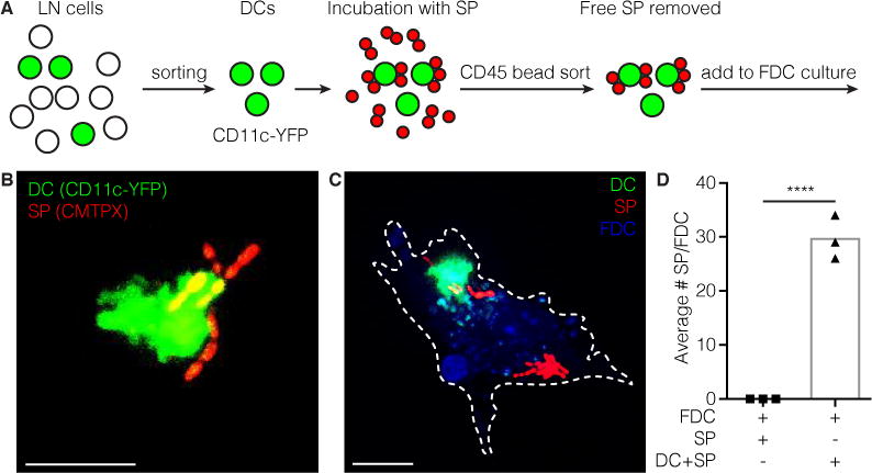
Figure 4. DCs can transfer SP directly to FDCs.

a | Schematic of experimental setup. LN single cell suspension from CD11c-YFP mouse is made. YFP positive DCs are FACS sorted. Sorted DCs are incubated with SP in the presence of complement components. Then DCs are sorted again using CD45 magnetic beads to remove unbound SP. DCs carrying SP are added to FDC cultures isolated 3 days prior. b | Sorted CD11c-YFP DC multiple carrying SP (CMTPX) on its surface. Scale bar 10 μm. c | FDC (blue) loaded with SP (CMTPX) and a DC (YFP) transferring SP. Scale bar 10 μm. d | Quantification of SP transferred to FDCs through DCs. **** p<0.0001, Students t-test.
