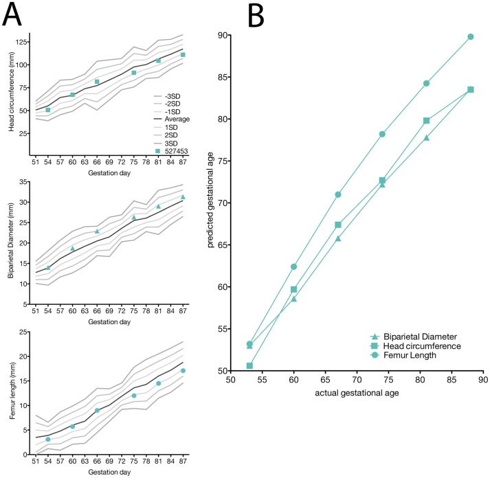
Fig 5. Fetal growth measured by ultrasonography.
(A) Head circumference (HC), biparietal diameter (BPD), and femur length (FL) were measured in weekly ultrasounds. All measurements are depicted as millimeters (mm). The solid lines were derived from reference ranges from Tarantal et al. 2005 to show the mean (black lines) and one, two, and three standard deviations from the mean (grey lines). The HC, BPD, and FL were then plotted along these reference ranges to observe any deviations from the mean. (B) The pGA is plotted against the aGA (based on gestational age estimated from breeding and menstrual history). The pGA is shown separately for each measurement: BPD (triangle), HC (square), and FL (circle).

