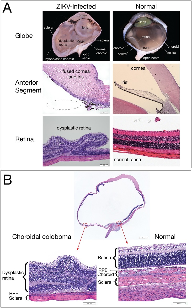
Fig 8. Fetal ocular pathology.
(A) The left panels contain images of the ZIKV-infected eye, and the right panels show normal features from a control gestational day 155 male rhesus macaque fetus for comparison. The globe of the ZIKV-infected fetus shows a hypoplastic choroid and dysplastic retina compared to the normal eye. The irregular shape of the eye in the ZIKV-infected globe is a processing artifact. The anterior segment image of the ZIKV-infected fetus shows that the iris is fused to the posterior cornea (black arrow heads), suggesting anterior segment dysgenesis; the dotted line shows where the iris would be in normal ocular development. The ZIKV-infected eye presents marked retinal dysplasia, characterized by retinal folding and loss of normal retinal organization when compared with the normal retina in the control image on the right. (B) A choroidal coloboma was identified on the ventral aspect of the globe (left image); the choroid had normal development on the dorsal aspect of the same globe (right image). The retina, retinal pigment epithelium (RPE), choroid (if present), and sclera are labeled with the left image demonstrating an absence of choroid.

