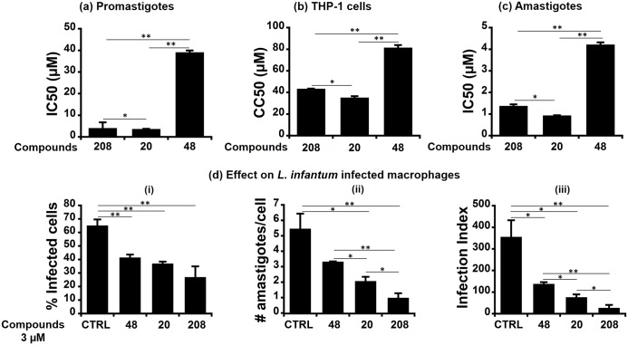
Fig 6. Hits 208, 20 and 48 impaired promastigote viability and amastigote survival in a dose-dependent manner.
(a) IC50 measured on promastigote viability: Stationary phase parasites were plated in 96-well plates at a final parasite density of 5x10⁶ parasites/ml, incubated for 24h in the presence of different concentrations of the studied compounds and counted. IC50 values were determined and plotted here for each compound. (b) CC50 measured on THP-1 derived macrophages: The cells were seeded in 96-well plates (50,000 cells/well), treated with serial concentrations of each compound for 24h, and counted. CC50 values were determined and plotted. (c) IC50 measured on intracellular amastigotes: THP-1 derived macrophages were infected with L. infantum strain at MOI of 10:1 for 24h. Upon cell washing to eliminate residual extracellular parasites, they were further incubated for 24h in presence of different compound concentrations. Number of intracellular amastigotes and infected cells were counted after Giemsa staining and IC50 values were determined and plotted. (d) Effect of the compounds on L. infantum infected cells at a concentration of 3 μM: The panel illustrates the percentage of infected THP-1 cells (i), the number of parasites per infected THP-1 (ii), and the infection index (iii). All results are shown as the mean ± SD of three independent experiments also done in technical triplicates. Statistical differences were analyzed with Student’s t-test ((* p < 0.05) or (** p < 0.01)).

