
Figure 2b:
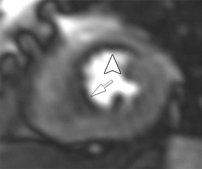
Stress perfusion defects in the left anterior descending coronary artery territory in a 74-year-old woman. Representative true-positive (a) CT perfusion and (b) MR perfusion images show a myocardial perfusion deficit in the anteroseptal wall. Note the anterior deficit (arrowhead) and the septal part (arrow). For visualization of the CT perfusion deficit, a short-axis screen shot with 8-mm section thickness is shown in the rainbow red preset. (c) ICA shows 90% stenosis (*) in the left anterior descending coronary artery. (d) SPECTimage shows a matching perfusion deficit in the anterior wall (arrowhead) and a less pronounced deficit in the septum (arrow).