
Figure 2a.
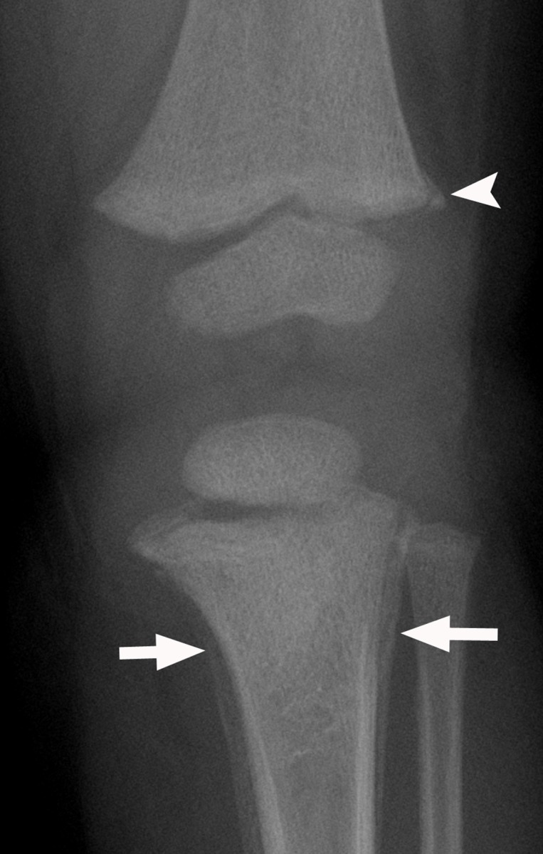
Anchoring bias confounded by confirmation bias. A 17-month-old girl presented with 1.5 months of intermittent leg pain and the inability to walk and reversion to crawling during the past month. (a) Frontal radiograph of the proximal left tibial metaphysis shows periosteal reactions (arrows), which were suspicious for malignancy. A calcific fragment (arrowhead) adjacent to the distal left femoral metaphysis was described as a likely developmental variant spur rather than a classic metaphyseal lesion, which would raise concern for nonaccidental trauma. Magnetic resonance (MR) imaging was recommended. (b) Coronal gadolinium-enhanced fat-saturated spoiled gradient-echo T1-weighted MR image shows periosteal enhancement (arrowheads) in the proximal left tibia. Absence of bone marrow edema or enhancement in the distal left femur was reassuring that the finding was a normal variant and not a sequel of trauma. (c) Axial CT image of the proximal left tibia shows a guided biopsy. Results of the biopsy helped confirm a healing fracture. In this case, the radiologist continued to pursue imaging and intervention that would support the initial diagnosis of malignancy.