
Figure 4b.
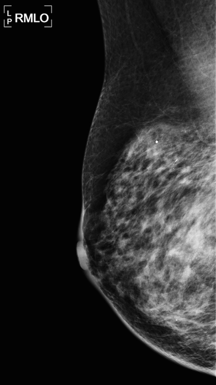
Satisfaction of report. A 65-year-old woman, with a history of bilateral lumpectomy followed by left mastectomy for disease recurrence, presented for right diagnostic mammography, per lumpectomy protocol. The patient denied having a palpable lump, pain, or nipple discharge. (a) Right mediolateral oblique (MLO) mammogram shows an area of increased density (arrow) at the lumpectomy site. (b, c) Right MLO mammograms from previous imaging studies from the past 2 years show the area of increased density, which was described in the previous reports as scarring and classified as Breast Imaging Reporting and Data System (BI-RADS) category 2. (d) MLO right mammogram from a remote prior imaging study shows the original lumpectomy scar, which helped confirm that the most recent mammogram (a) represents a developing asymmetry at the lumpectomy site. (e) Antiradial US image shows a shadowing mass (arrow), which was palpable at clinical examination. Pathologic analysis helped confirmed recurrent breast carcinoma.