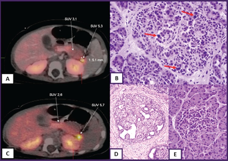
Figure 3. 18F-fluoro-L-dihydroxyphenylalanine (18F-DOPA)-positron emission tomography/computed tomography scan images of focal congenital hyperinsulinism (A and C), histological figure of diffuse (B) and focal (D) disease and normal pancreas islet cell (E). Standardized uptake value (SUV) 5.3 and SUV 5.7 indicate focal uptake of 18F-DOPA, red arrows show large nuclei of β-cell in diffuse disease.

