Figure 1.
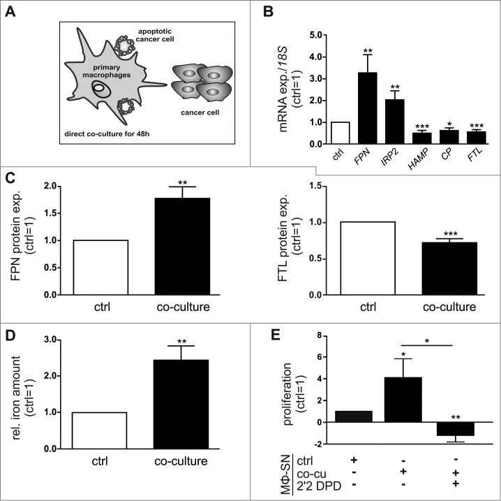
Macrophages acquire an alternatively activated, iron-release phenotype in vitro. (A) Schematic overview of the direct co-culture model. Primary human MΦ were co-cultured with MCF-7 breast cancer cells for 48 h, unless indicated otherwise. (B) mRNA expression of iron-regulated genes (FPN, IRP2, HAMP, CP, FTL) in MΦ after co-culture (solid bars) was analyzed by qPCR and normalized to 18 S expression (n = 9-20). (C) Protein expression of FPN (surface, left panel) and FTL (intracellular, right panel) in MΦ after co-culture (solid bars) was determined by flow cytometry and normalized to the respective expression in naïve control MΦ (ctrl, open bars) (n = 9). (D) Iron amount in the supernatant of MΦ after co-culture (solid bar) was quantified by AAS and normalized to the supernatants of naïve control MΦ (ctrl, open bar) (n = 5). (E) Proliferation of MCF-7 cells upon stimulation with supernatants from MΦ (MΦ-SN) co-cultured with tumor cells (co-cu) was determined on an xCELLigence instrument as an increase in the impedance (slope 1/h) and is given relative to MCF-7 cells treated with supernatants from naïve control MΦ (ctrl) (n = 5). Data are means ± SEM from independent experiments, *p < 0.05, **p < 0.01, ***p < 0.001 vs. the respective controls unless indicated otherwise.

