
Figure 1.
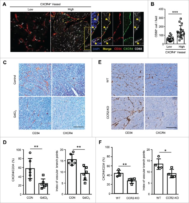
Tumor-infiltrating Mo/Mϕ regulate vascular CXCR4 expression in HCC tumor tissues. (A) The staining of CD34+ vascular (red), CXCR4+ (green) and CD68+ monocytes/macrophages (gray) in HCC tissues was examined by confocal microscopy. DAPI staining appears blue. Scale bar = 50 μm. (B) Patients were divided into two groups according to the median value of the intra-tumoral CXCR4+ vascular densities (n = 30). The infiltration of Mo/Mϕ between groups are shown. (C−F) Orthotopic HCC models derived from Hepa1-6 cells were established in C57 BL/6 (C and D) or CCR2-KO (E and F) mice. Immunohistochemical staining for CXCR4 and CD34 was performed on serial tissue sections of the mouse tumor allografts. Scale bar = 100 μm. The vascular CXCR4 density and the index of the vascular branch points were assessed (D and F). WT, wild type; CCR2-KO, CCR2 knock-out mice. Values given in B, D, and F represent the means ± SEM. * P < 0.05; ** P < 0.01; *** P < 0.001.