
Figure 2.
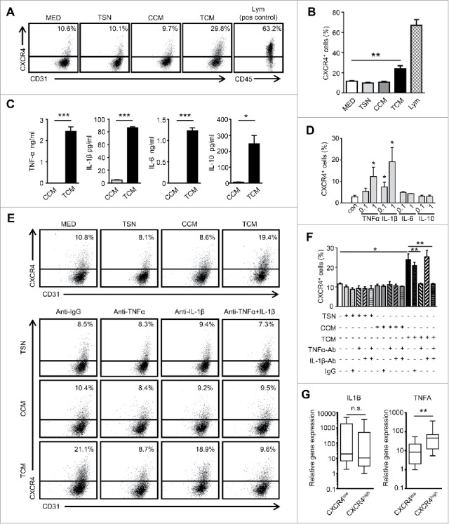
Conditioned medium from tumor-activated Mo/Mϕ induce CXCR4 expression on ECs. (A and B) HUVECs were cultured for 24 hrs in conditioned medium from tumor cells (TSN), control monocytes (CCM) or medium from TSN-exposed monocytes (TCM). Peripheral blood lymphocytes were used as the positive control for CXCR4 staining. (C) The levels of cytokines in CCM or TCM were determined by ELISA. (D) HUVECs were incubated for 24 hrs with rhTNF-α, rhIL-1β, rhIL-6, or rhIL-10 (ng/ml) at the indicated concentrations. (E and F) The HUVECs were cultured in TSN, CCM or TCM and mAb against TNF-α (1 μg/mL) or IL-1β (10 μg/mL), combined, or the control Ab (IgG1, 10 μg/mL). The percentages of HUVECs expressing CXCR4 were determined by FACS. (G) The relative expression of the indicated gene mRNA levels in the tumor tissues from HCC patients (n = 25) were analyzed by qPCR. All data shown are representative of at least three independent experiments. Lym, Peripheral blood lymphocytes. Error bar, SEM; n.s., not significant; * P < 0.05; ** P < 0.01; *** P < 0.001.