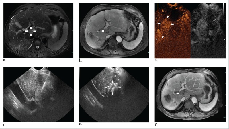
Figure 1.

A 63-year-old man with hepatocellular carcinoma. Preoperative MR and CEUS images showed a mass of 2.2*1.7 cm in size in the caudate lobe (a, b and c) (white arrows). EUS suggested laser fiber inserted into the tumor (d) and then total enhancement of the lesion (e) (white arrows). One year later, substance phase MR image showed that the mass has a complete response (f) (white arrows).
