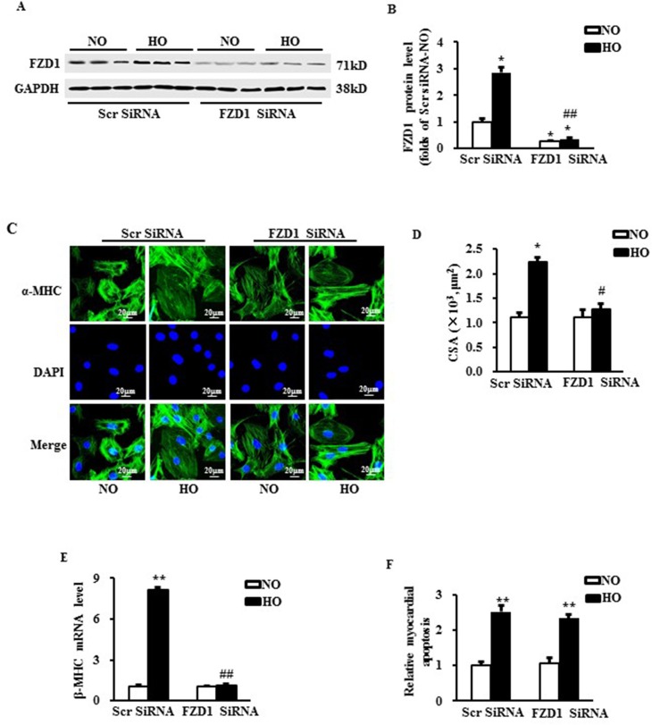
Figure 2. Deficiency of FZD1 alleviated hypoxia-induced myocardial hypertrophy in vitro.
(A) Representative Western blots of FZD1 in neonatal cardiomyocytes (NRCMs) after transduced with indicated SiRNA and treated with hypoxia. N = 5 independent experiments. (B) Quantitative results of the protein expression of FZD1in NRCMs. (C) The representative images of NRCMs stained by α- myosin heavy chain (α-MHC). (D) The quantification data of NRCM surface area (CSA). (E) mRNA levels of hypertrophic markers, β-myosin heavy chain (β-MHC) in NRCMs measured by real-time PCR. HPRT was served as internal control. (F) The myocardial apoptosis expressed as the ratio of TUNEL-positive nuclei over DAPI-stained nuclei. N = 5 independent experiments. GAPDH was served as internal control in Western blot. NO, normoxia. HO, hypoxia. All data are expressed as mean ± S.E.M. *P < 0.05 compared with NO + Scr SiRNA, #P < 0.05 compared with HO + Scr SiRNA.

