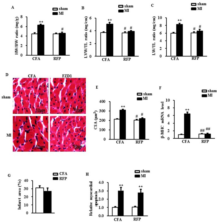
Figure 4. RFP attenuated cardiac hypertrophy and rescued cardiac dysfunction after MI.
(A) Heart weight/ body weight ratio (HW/BW), (B) Left ventricular weight/ tibia length ratio (LVW/TL) and (C) Lung weight/ tibia length ratio (LW/TL) of mice treated by indicated surgery and treatments. (D) Representative images of histological sections of the mouse LVs were stained with hematoxylin-eosin (H&E) one week after MI or sham surgery. Scale bar: 50 μm. (E) Quantitative results of the cross sectional area (CSA) of mouse cardiomyocytes quantified by using an image analysis system. (F) Real-time PCR analysis of β-MHC in mouse LVs after MI. HPRT was served as internal control. (G) The relative infarcted area of mouse left ventricles. (H) the relative ratio of myocardial apoptosis in the infarct border zone of the mouse left ventricles. N = 5 per experimental group. CFA, Complete Freund’s adjuvant. N = 5 independent experiments. All data are expressed as mean ± S.E.M. **P < 0.01 compared with sham + CFA, #P < 0.05, ##P <0.01 compared with MI + CFA.

