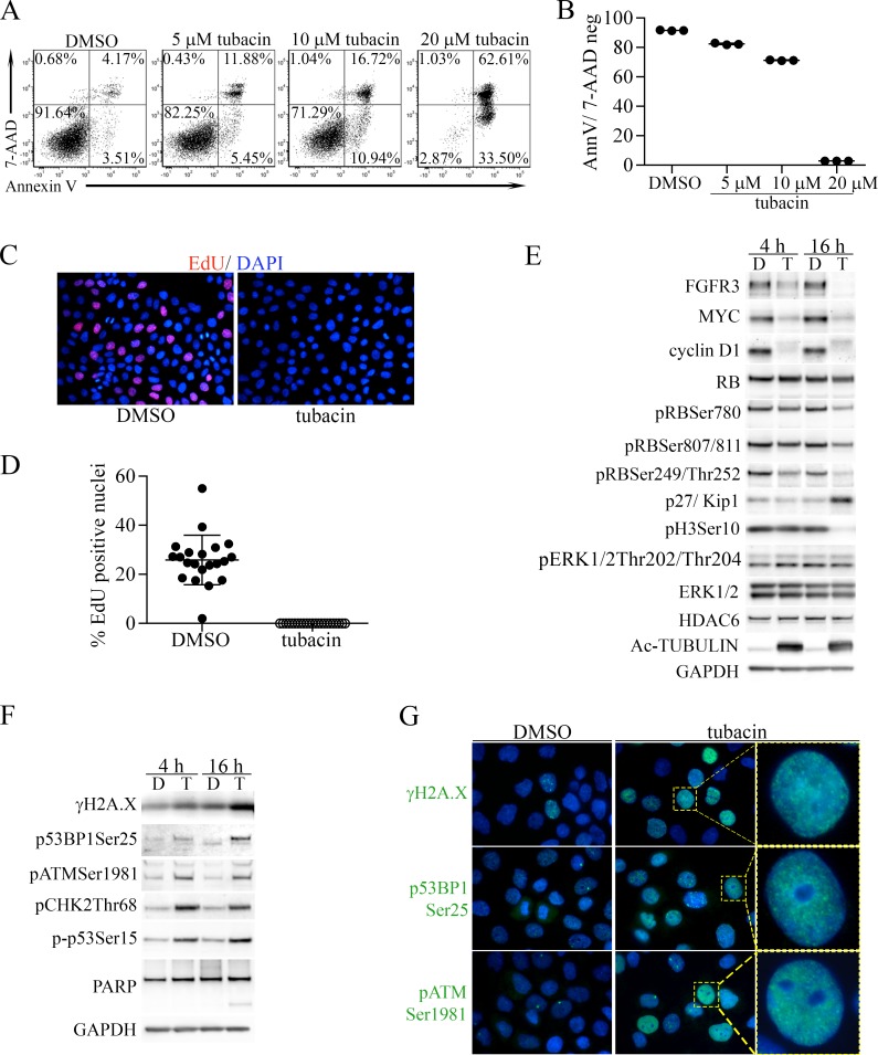
Figure 6. Induction of apoptosis and proliferative arrest by tubacin in RT112 bladder cancer cells is associated with downregulation of FGFR3, MYC, cyclin D1 and induction of DNA damage.
(A) Representative FACS analysis of 7-AAD- and Annexin V-stained RT112 cells treated with DMSO vehicle or the indicated concentrations of tubacin for 48 hours. (B) Dot plot of results from three experiments examining apoptosis at the indicated tubacin concentrations. (C) Representative image of staining for EdU (red) incorporation in DNA by RT112 cells treated with DMSO or tubacin for 16 hours. Cells were co-stained with DAPI (blue) to mark nuclei. (D) Dot plot analysis showing the percentage of EdU positive nuclei per image (N) (DMSO, N = 21; tubacin, N = 23). (E) Immunoblots for FGFR3 (FGFR3-TACC) and the indicated proteins and phospho-proteins associated with cell proliferation and cyclin D1 activity in RT112 cells treated with DMSO (D) or 20 µM tubacin (T) for 4 or 16 hours. (F) Immunoblots for the indicated proteins associated with DNA damage signaling, DNA repair and apoptosis from RT112 cells treated with DMSO (D) or 20 µM tubacin (T) for 4 or 16 hours. (G) Immunofluorescent staining of the indicated proteins (green) in RT112 cells treated for 16 hours with DMSO or 20 µM tubacin. DAPI stained nuclei are blue. Higher magnification images of the indicated cells (yellow boxes) showing punctate staining patterns are shown at right.

