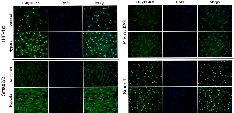
Figure 2. HIF-1α, Smad2/3, p-Smad2/3 and Smad4 was enhanced following treatment with 1% hypoxia.
The protein expression and intracellular localization of HIF-1α, Smad2/3, p-Smad2/3 and Smad4 were detected by immunofluorescence staining in HFFs under normoxia or 24 h of hypoxia (1% O2). HIF-1α and Smad4 localized mainly in the nucleus and Smad2/3 mainly in cytoplasm.

