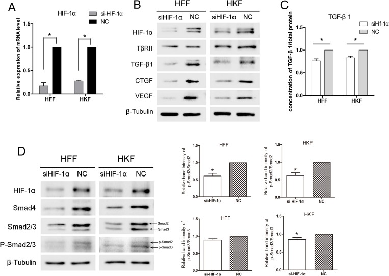
Figure 3. siHIF-1α inhibited TGF-β/Smad signaling in HFFs and HKFs.
(A) qRT-PCR showing clear knockdown of HIF-1α by siHIF-1α transfection for 48 h. (B, D) 48 h after transfection, HFFs and HKFs were transferred to a 1% O2 hypoxia incubator for 24 h, and siHIF-1α inhibited HIF-1α, TGF-β1, Smad4, Smad2/3, p-Smad2/3, CTGF and VEGF levels. The histogram shows the protein band intensity ratio of p-Smad2 to Smad2 and p-Smad3 to Smad3. (C) Before transferring cells to the hypoxia incubator, we replaced the culture media with serum-free media, and both the siHIF-1α and NC groups were treated with 1% O2 for 12 h. The level of secreted TGF-β1 in the serum-free media was then measured.

