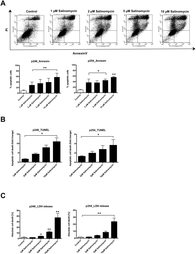
Figure 2. Treatment with Salinomycin induces apoptosis in murine CC cells.
(A) A total of 0.5 × 10⁶ p246 or p254 cells were seeded in six-well plates and grown until confluence following exposure to increasing concentrations of Salinomycin (1, 2, 5, and 10 µM) for 24 h. Detection of apoptosis was performed using AnnexinV-FITC and propidium iodide staining, and cells were analyzed by flow cytometry. Cell death was further determined by quantification of DNA fragmentation (B) and LDH release assay (C). Results are displayed as representative dot blots or as a summary of at least three independent experiments; *P < 0.05; **P < 0.001 compared with control.

