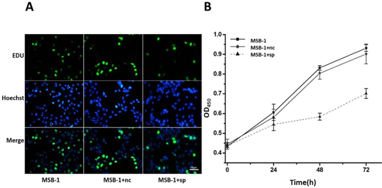
Figure 3. Effect of expression of MDV-1 miRNA sponge on proliferation of MSB-1 cells.
(A) miRNA sponge cells and control cells were incubated with EDU. After 24 h, EDU staining and DNA staining were used to observe the staining under fluorescence microscope. (B) The cells which were cultured in the incubator for 4 h were determined by OD value at 0 h. Then we determined OD at 24 h, 48 h, 72 h, respectively.

