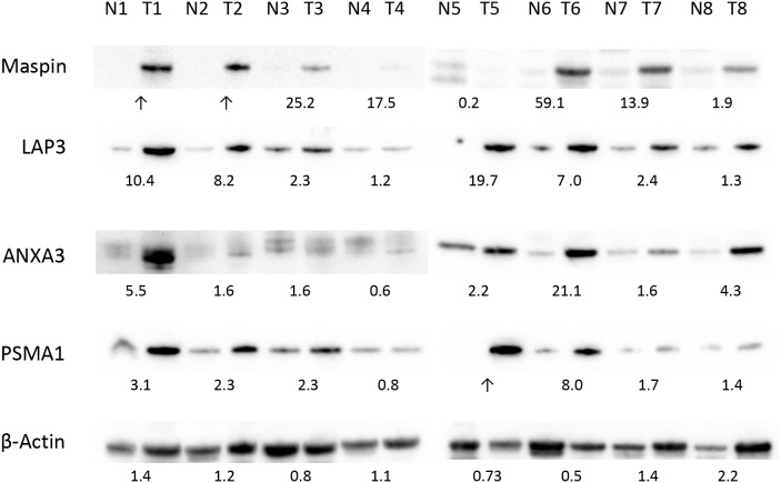
Figure 3. Differential expression of maspin, ANXA3, LAP3 and PSMA1 in colon cancer tissue validated by western blotting.
Protein extracts from 8 pairs of colon tumor (T1-8) and normal tissue (N1-8) were separated by SDS PAGE and blotted with commercial antibodies specific to each protein. Expression of β-actin in each tissue protein extract was used as a control. The numbers under each pair of blots represent the relative expression ratios, with ratios <1, =1, >1 indicating decreased, unchanged, or increased expression in tumor cancer, respectively.

