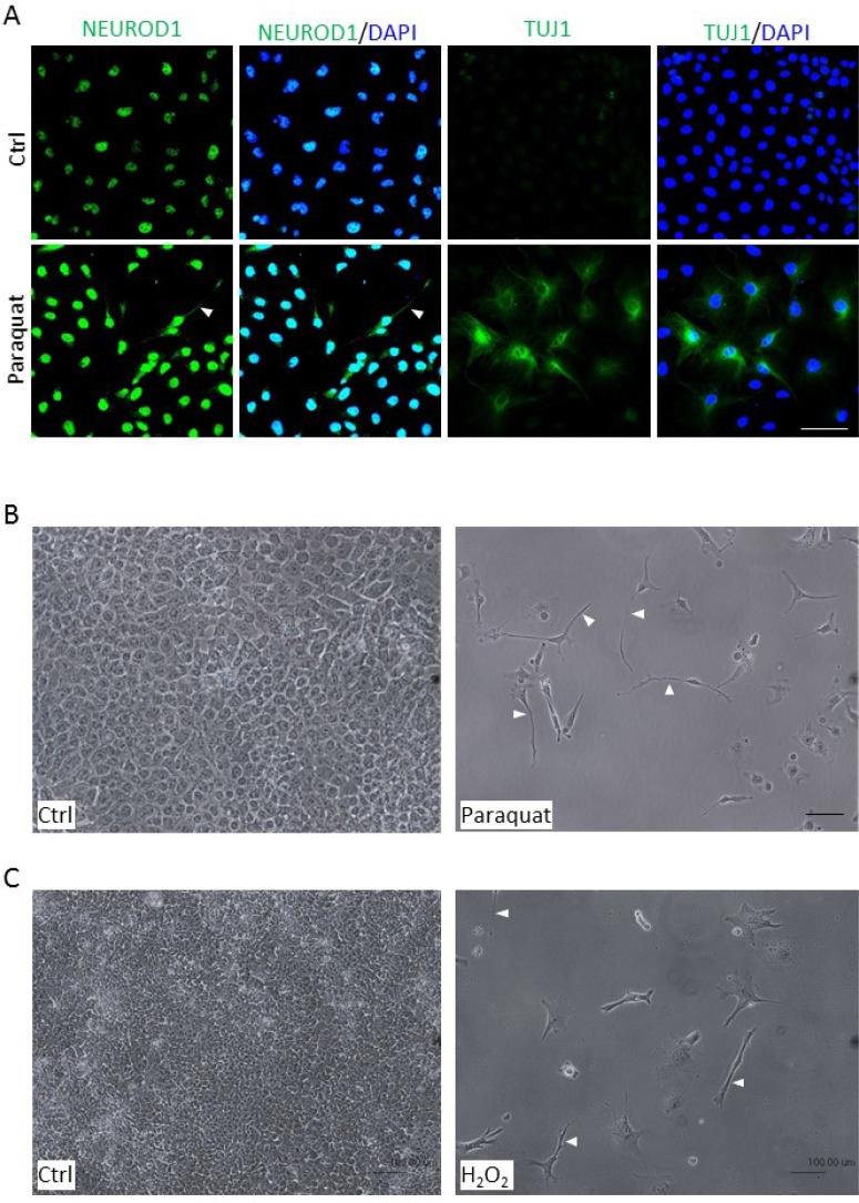
Figure 5. Enhanced ROS cause morphological changes in NT2 cells.
(A) Cells were treated with 25 μM paraquat for six days and immunostained with neuronal markers, NEUROD1 and TUJ1. The cells were counterstained with DAPI. The arrowhead denotes neurite-like cellular process. (B, C) Cells were treated with 100 μM paraquat (B) or 5 nM H2O2 (C) for six days, and the morphological changes were visualized by phase-contrast microscopy. The arrowheads point to elongated cellular processes. Scale bar: 100 μm.

