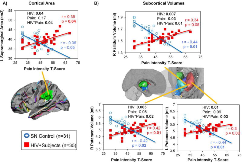
Figure 3. Group Differences in the Relationships between Pain Intensity and Brain Morphologic Measures in HIV+ Participants and SN Controls.
HIV+ individuals and SN controls showed opposite relationships between pain intensity and brain morphometry in the L supramarginal area, bilateral putamen volumes, and R pallidum volume. These graphs show that HIV individuals with higher pain intensity tended to have bigger cortical area in the L supramarginal region and larger subcortical volume in the R pallidum and bilateral putamen, while SN controls with higher pain intensity scores had smaller left supramarginal area, and smaller volumes in the right pallidum and bilateral putamen.

