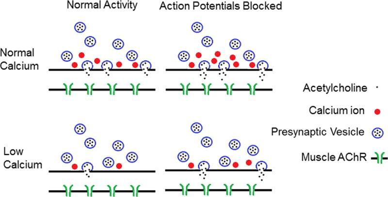
Figure 1.
Shown is a cartoon of homeostatic synaptic plasticity at the mouse NMJ triggered by prolonged blockage of nerve action potentials. In the top row, synaptic function is shown in solution containing normal external Ca. Following blockage of action potentials, there are two changes in synaptic function. The first is an increase in the release of acetylcholine (ACh) during fusion of individual synaptic vesicles (illustrated as increased ACh in the synaptic cleft). This is likely responsible for the increase in the amplitude of spontaneous synaptic currents (miniature endplate currents (MEPCs)). The second change is an increase in entry of Ca into the presynaptic terminal during an action potential. However, when extracellular Ca is normal, this has no significant effect on the number of synaptic vesicles released (quantal content (QC)), as each releasable vesicle is already released with each action potential. The bottom row shows the situation when extracellular Ca is lowered. In this case, Ca entry during the action potential limits QC. When Ca entry is increased following blockage of action potentials, even a small increase in Ca entry triggers a significant increase in QC. In addition, the increase in ACh release from each vesicle also occurs. This combines with the increase in QC to cause a dramatic increase in synaptic strength. This is illustrated in the cartoon as an increase from two to eight ACh molecules in the synaptic cleft. AChR, acetylcholine receptor.

