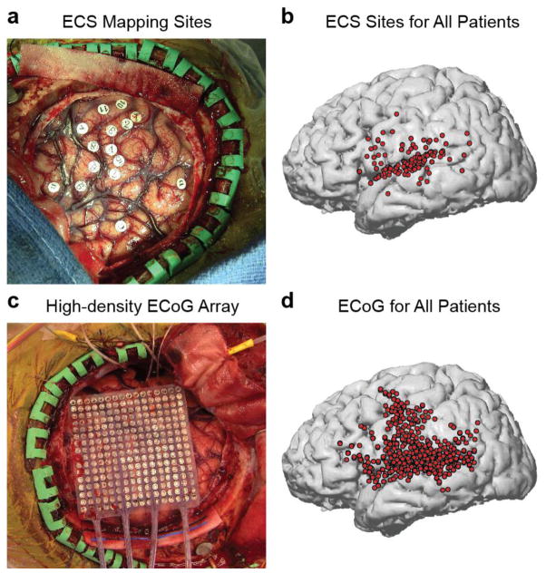
Figure 1. Two complementary methods for examining the cortical networks involved in verbal repetition.
(a) Example intraoperative photograph showing exposed craniotomy and markers where electrocortical stimulation was performed. (b) Reconstructed brain showing all positive stimulation sites across 47 patients, covering the major peri-Sylvian regions hypothesized to be involved in verbal repetition. (c) Example intraoperative photograph showing exposed craniotomy and high-density 256-channel ECoG grid covering peri-Sylvian cortex. (d) Reconstructed brain showing all electrode locations included in the ECoG analyses for 8 patients.

