
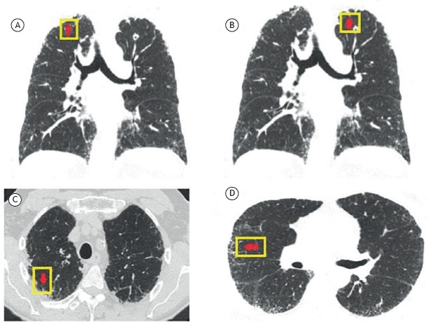
Figure 1. Coronal (A) and axial (B, C, and D) CT scans of the chest showing intense pleural and subpleural fibrosis, as well as septal thickening, predominantly located in the upper lobes and a spiculated nodule in the left upper lobe.
Official websites use .gov
A
.gov website belongs to an official
government organization in the United States.
Secure .gov websites use HTTPS
A lock (
) or https:// means you've safely
connected to the .gov website. Share sensitive
information only on official, secure websites.