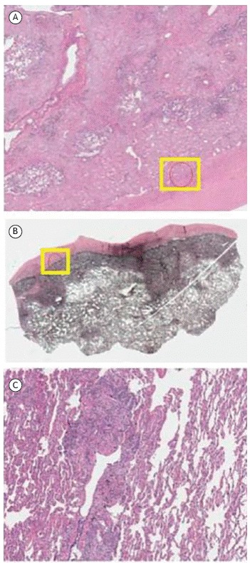
Figure 3. Upper lobe explant histological findings. In A and B, presence of fibrous thickening of the visceral pleura and subpleural lung parenchyma, with little dense collagen deposition and abundant elastic fibers that are evident on Verhoeff staining. In C, signs of small airways disease, either by obliteration or by distortion of the bronchial wall, accompanied by malformed granulomas and multinucleated giant cells containing cholesterol crystals, located interstitially or intra-alveolarly.

