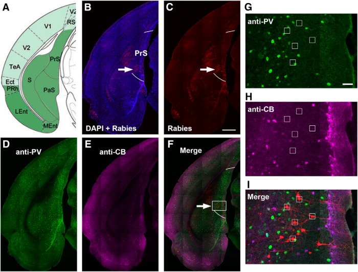
Figure 5.
Rabies-labeled CA1-projecting neurons in immunochemically delineated presubiculum are mostly excitatory neurons. A, The mouse atlas image shows the anatomic location of the presubiculum (PrS). B, C, A brain section image corresponding to the atlas image. Rabies-labeled neurons appear in the presubiculum (indicated by the white arrow) after CA1 virus injection. DAPI staining is blue. Two white lines delineate the region of PrS per PV and CB immunochemical staining. The scale bar (500 μm) applies to B–F. D–F, Images of parvalbumin (PV) immunostaining (green, D), calbindin-D28k (CB) immunostaining (magenta, E), and a merged image (F). White arrow points to the region with rabies-labeled neurons, which has strong PV immunoreactivity and weak calbindin immunoreactivity. The PV and CB immunoreactivity features allow for delineation of PrS (Fujimaru and Kosaka, 1996; Fujise et al., 1995). G–I, Enlarged images of the white box region in F show PV staining (G), CB staining (H), and rabies-labeled neurons (red) in the merged image (I). Small white squares locate rabies-labeled neurons that are not positive for PV or CB staining. The scale bar (100 μm) in G applies to G–I.

