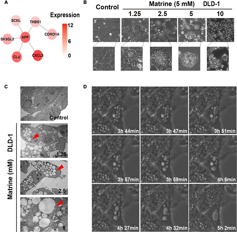
FIGURE 4.

Matrine induces vesicle formation, endocytosis and macropinocytosis in a time-and concentration-dependent manner. (A) The endocytosis-associated network module of matrine with expression in DLD-1. (B) Phase-contrast microscopy of the matrine-treated DLD-1 cells for 12 h shows extensive accumulation of cytoplasmic vacuoles and cell detachment in a concentration-dependent manner. (C) TEM images of matrine-treated DLD-1 cells at different concentrations for 12 h. Red arrow points at cytoplasmic vacuoles. Bar, 10 μM. (D) Live imaging of DLD-1 cells (5 mM matrine). Images collected at times of major phenotypic changes.
