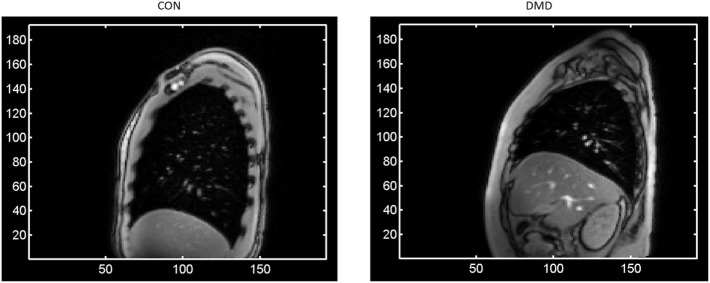
Figure 1.
A single frame of the right sagittal dynamic Magnetic Resonance Imaging data for two representative subjects at baseline—one from each group [left: a control; right: a Duchenne Muscular Dystrophy (DMD) patient]—showing at full lung capacity the much larger lung cross-sectional area for the control compared to the DMD subject, as well as the typical levels of signal-to-noise and the observed image features such as the contrast at the periphery of the lung. The axes represent pixel coordinates, and the image field-of-view was the same in each case.

