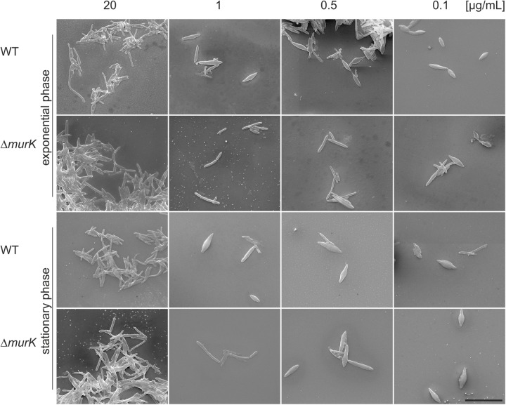
FIGURE 6.
SEM micrographs of T. forsythia wild-type (WT) and ΔTf_murK::erm (ΔmurK) mutant cells when grown under full MurNAc supplementation (first row) and under MurNAc limiting conditions (rows two to four). Media were supplemented with 20 μg/ml (full supplementation), 1 μg/ml, 0.5 μg/ml, and 0.1 μg/ml of MurNAc. The ΔmurK mutant is more tolerant toward MurNAc limitation as can be seen from transition from rod-shaped to fusiform cells occurring only at 0.1 μg/ml MurNAc, with this effect being more profound in the exponential growth phase. Scale bar, 10 μm.

