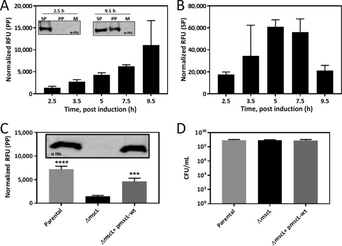
FIG 3 .
Analysis of recombinant protein localization. (A) Localization of eGFP in the periplasm (PP) over time in BL21(DE3) and representative Western blot analyses (insets). (B) Localization of eGFP in the spheroplast (SP) over time in BL21(DE3). (C) Localization of eGFP in the periplasm of parental, MscL-deficient (ΔmscL), and restored (ΔmscL/pmscL-wt) E. coli K-12 cells at 16 h postinduction and representative Western blot analysis (inset). (D) CFU assay of parental, MscL-deficient (ΔmscL), and restored E. coli K-12 cells. eGFP fluorescence is shown as RFU normalized to cell density. Data are presented as the mean value ± the standard deviation of at least three biological replicates. *, P < 0.05; ***, P < 0.001; ****, P < 0.0001 (two-way ANOVA followed by Dunnett’s multiple-comparison test).

