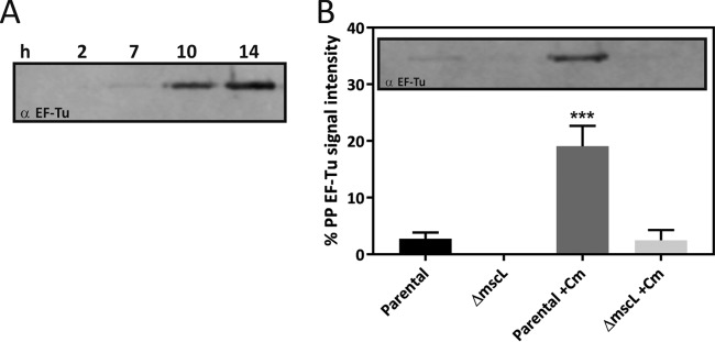
FIG 6 .
Excretion of cytoplasmic protein EF-Tu in the periplasm following translation stress. (A) Western blot analysis of EF-Tu localization in the periplasmic (PP) fraction from parental E. coli K-12 in the presence of Cm (0.01 mg/ml) over time. (B) Periplasmic localization of EF-Tu shown as a percentage of the total (periplasm [PP] plus spheroplast [SP]) protein, in parental and MscL-deficient E. coli K-12 cells in the absence or presence of Cm (0.01 mg/ml) after 10 h of treatment. A representative Western blot analysis is shown as an inset. Data in panel B are presented as the mean value ± the standard deviation of three biological replicates. ***, P < 0.001 (two-way ANOVA followed by Dunnett’s multiple-comparison test).

