FIG 1.

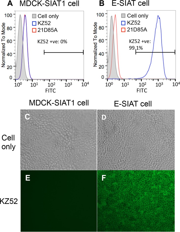
Stable expression of EBOV-GP on the surface of transduced MDCK-SIAT1 cells. Shown are FACS plots of MDCK-SIAT1 cells (A) and MDCK-SIAT1 cells transduced with full-length GP (E-SIAT) (B). Cells were stained with primary MAb KZ52 (anti-GP MAb) or 21D85A (anti-H5 MAb), followed by an FITC-linked anti-human secondary antibody. E-SIAT cells stably express GP at the cell surface up to at least the ninth passage, which can be detected with conformational MAb KZ52 specifically (B, blue). (C to F) Immunofluorescence pictures of MDCK-SIAT1 (C and E) and E-SIAT (D and F) cells stained with KZ52 and an FITC-linked anti-human antibody.
