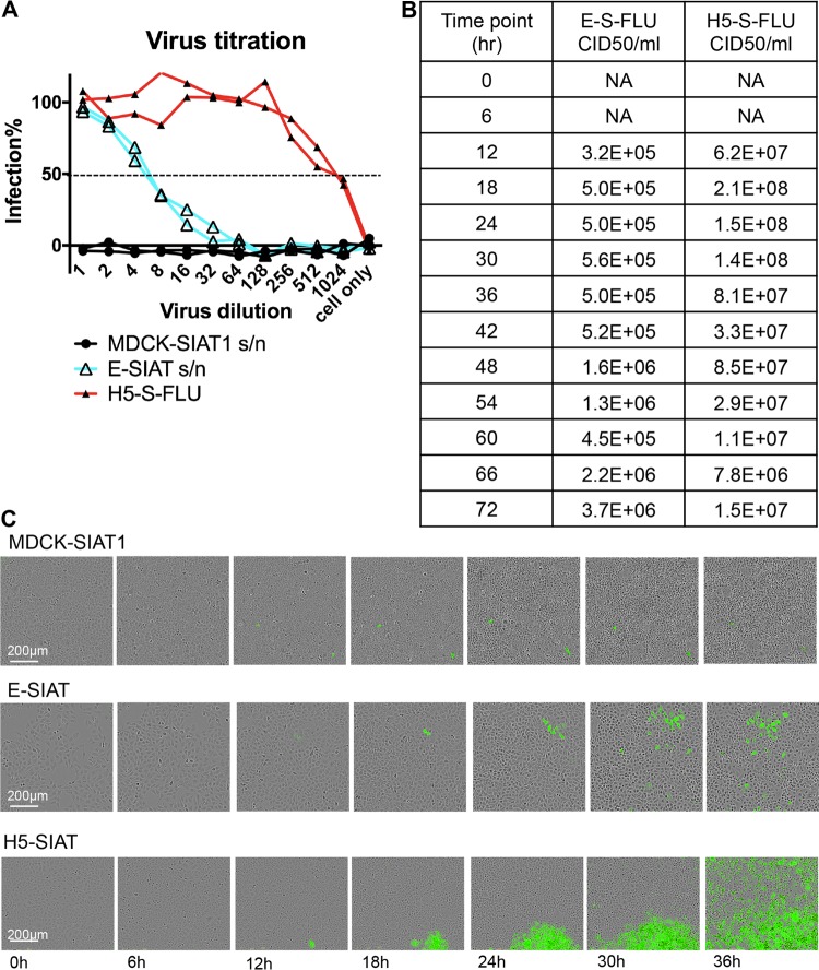
FIG 2.
Pseudotyping of S-FLU virus with EBOV-GP. (A) E-SIAT cells were seeded with 1 TCID50 of seed S-FLU virus per cell to generate EBOV-GP (cyan)- and H5 (red)-pseudotyped S-FLU viruses. Parental MDCK-SIAT1 cells were also included as a control. Culture supernatant (s/n) harvested after 48 h was titrated in comparison to the H5-S-FLU virus in 2-fold dilutions on MDCK-SIAT1 indicator cells for 24 h of infection. Infected cells expressing eGFP were detected and quantified with a CLARIOstar fluorescence plate reader. The undiluted E-S-FLU and H5-S-FLU viruses gave saturated infection, and the maximum fluorescence readout was used to calculate the percent infection at lower dilutions. (B) E-SIAT and H5-SIAT cells were seeded with 1 TCID50 of seed S-FLU virus per cell. Culture supernatant harvested at different time points postseeding were titrated on MDCK-SIAT1 cells. The EC50 was calculated by linear interpolation. The CID50/ml was calculated from the EC50 dilution and the number of cells per well (3 × 10⁴). (C) A total of 100 TCID50 of seed S-FLU virus was added to 3 × 10⁴ MDCK-SIAT1, E-SIAT, or H5-SIAT cells. Live images of representative plaques were taken by IncuCyte every 3 h. E-SIAT cells support the formation of small diffuse plaques, unlike the dense plaques formed on H5-SIAT cells.

