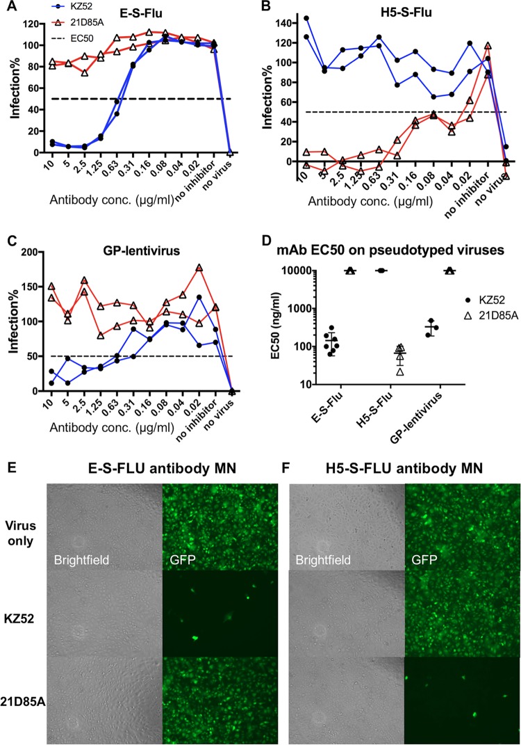
FIG 4.
Antibody neutralization of pseudotyped E-S-FLU virus, H5-S-FLU virus, and EBOV-GP-pseudotyped lentivirus. Viruses were preincubated for 2 h with antibody KZ52 (anti-GP) or 21D85A (anti-H5) before MDCK-SIAT1 indicator cells were added. Antibodies were titrated in 2-fold dilutions from 10 μg/ml. After 24 h, infection was quantified by measuring eGFP expression (influenza virus) or luminescence (lentivirus). Titration curves are shown in panels A to C. A summary of the EC50s of MAbs KZ52 and 21D85A for the three pseudotyped viruses is shown in panel D. Multiple EC50s from repeat experiments are shown with the geometric means and 95% CIs. A value of 10,000 ng/ml was assigned to antibodies that did not neutralize. (E and F) Microscopy (10× objective) of assays showing the specificity of MN by antibodies. For “Virus only,” the E-S-FLU and H5-S-FLU viruses were added directly to MDCK-SIAT1 cells. Infected cells express eGFP, shown as green cells. In the MN assay, virus was preincubated with 10 μg/ml MAb KZ52 (anti-GP) or 21D85A (anti-H5) before the infection of MDCK-SIAT1 cells. Inhibition is visualized as a significantly reduced number of green fluorescent cells.

