FIG 5.
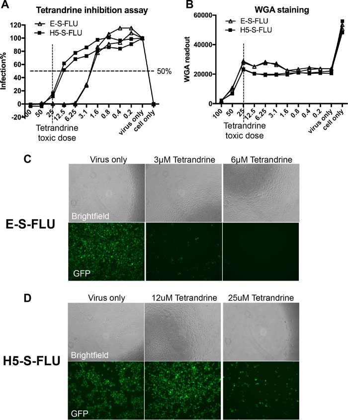
Tetrandrine inhibition assay with the E-S-FLU and H5-S-FLU viruses. Tetrandrine was titrated from 100 to 0.2 μM in the drug inhibition assay against the E-S-FLU and H5-S-FLU viruses. Cells were preincubated with tetrandrine for 3 h and infected with virus for 24 h. (A) Percent infection was quantified by eGFP expression measured with a fluorescence plate reader. (B) WGA was added to stain cell membrane sialic acid and N-acetylglucosaminyl residues as an estimation of the number of cells remaining in each well after fixation and washing. Toxicity reduces the WGA fluorescence as cells detach from the plastic. Note that the S-FLU virus expresses neuraminidase, which partially reduces WGA binding between “cell only” and “virus only” in panel B. Neutralization of the S-FLU virus is associated with some increase in WGA binding as neuraminidase expression is reduced. (C and D) Microscopy of tetrandrine inhibition assays. Note that the toxicity of tetrandrine at 25 μM resulted in a reduction in the number of cells in the bright-field channel.

