FIG 3.
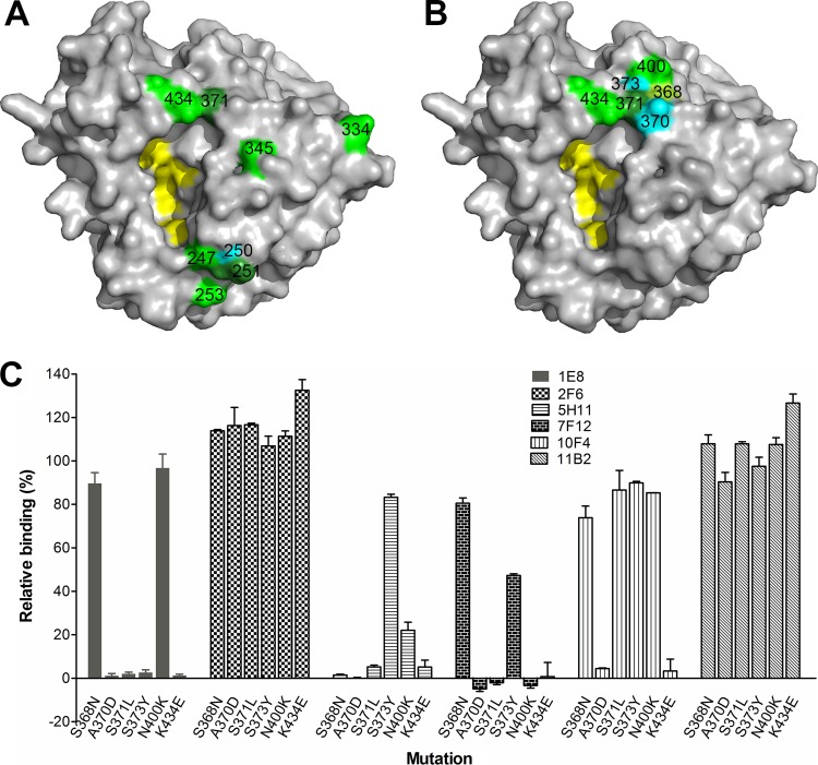
Residues on N9 that are critical for MAb binding. (A) Locations of the eight key residues (identified through escape mutant selection) on an N9 monomer (PDB accession number 4MWW). The image was generated with PyMOL software (Delano Scientific). Different colors were used to differentiate neighboring residues when necessary. (B) Residues in the 370, 400, and 430 loops that constitute the HB site on N9 that endow it with its ability to agglutinate red blood cells. Residues 118, 119, 224, 227, and 406 are highlighted in yellow in both panels A and B to depict the location of the NA active center. (C) Impact of amino acid mutations within the HB site of AH/13 NA on the binding by MAbs 1E8, 2F6, 5H11, 7F12, 10F4, and 11B2. Binding was measured by cell-based ELISA, with the signals expressed as percentages of those obtained with wt AH/13 NA. Shown are the means plus standard deviations from two independent assays run in duplicate wells.

