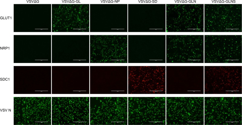
FIG 2.
Expression of HTLV-1 receptor molecule(s) on the rVSV-infected target cell surface. The surface expression of GLUT1, NRP1, or SDC1 protein after VSV infection was confirmed by IF. VSV-permissive BHK-21 cells were infected with each G-complemented rVSV (VSVΔG, VSVΔG-GL, VSVΔG-NP, VSVΔG-SD, VSVΔG-GLN, or VSVΔG-GLNS) at an MOI of 0.1. After 3 days of culture, target molecules expressed by the viral genome were stained with FITC-conjugated (GLUT1 and NRP1) or PE-conjugated (SDC1) specific antibodies without any fixation or permeabilization. Intracellular staining of VSV N protein was performed to detect VSV-infected cells, using mouse anti-VSV N MAb followed by FITC-conjugated anti-mouse IgG. The stained cells were observed by fluorescence microscopy and photographed at a constant magnification. Scale bars in panels represent 400 μm.

