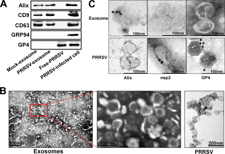
FIG 1.
Isolation and characterization of exosomes from PRRSV-infected cells. (A) Purified exosomes derived from mock- or PRRSV-infected cells were analyzed on Western blots probed with antibody directed against Alix, CD9, CD63, GRP94, or GP4. The PRRSV-infected cells and purified virions (free PRRSV) were used as controls. (B) Transmission electron microscopy observations of negatively stained purified exosomes from PRRSV-infected cells and free PRRSV. Purified exosomes with both higher and lower magnification are shown. (C) Immunoelectron microscopy images of purified exosomes or virions from PRRSV-infected cells. Immunogold labeling (10-nm gold particles) was performed with antibodies against exosome marker protein Alix and viral proteins nsp2 and GP4, respectively.

