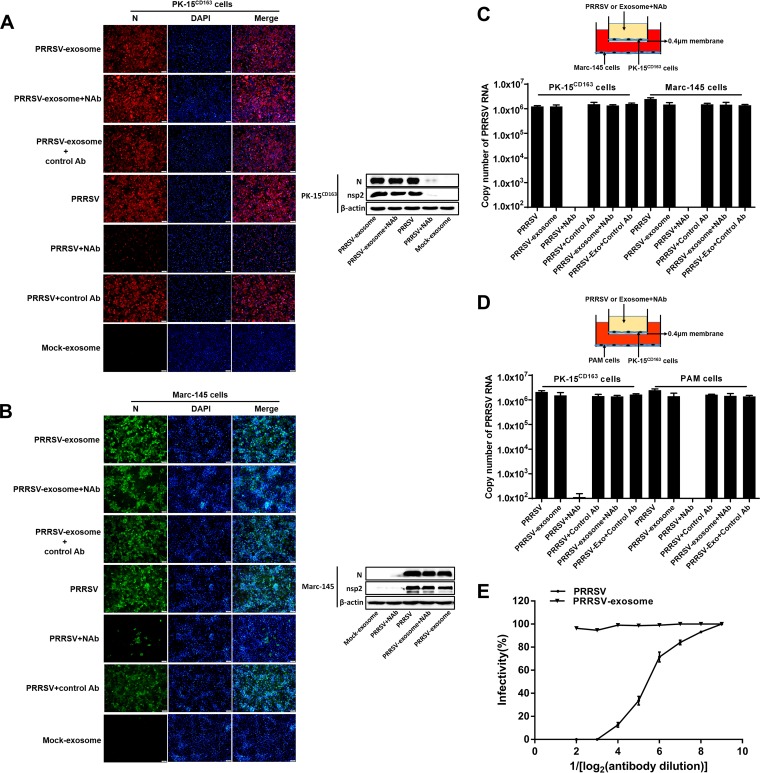
FIG 5.
Exosome-mediated PRRSV infection is not blocked by PRRSV-specific neutralizing antibodies (NAbs). (A, B) Purified PRRSV-positive exosomes or free PRRSV virions were incubated with PRRSV-specific NAbs for 1 h. Then, PK-15CD163 (A) or Marc-145 (B) cells were exposed to the antibody-treated exosomes or virus for 3 h. The exosomes or viruses were washed off, and the medium was replaced with fresh maintenance medium for a further 36 h. Immunofluorescent staining was performed with MAb against PRRSV N protein. Western blotting was performed with MAbs against PRRSV N and nsp2 proteins. (C) Schematic diagram of the Transwell system used to coculture PK-15CD163 and Marc-145 cells. PK-15CD163 cells in the upper chamber were incubated with purified PRRSV-positive exosomes or infected with PRRSV pretreated with PRRSV-specific NAbs (neutralization titer is 1:16) for 1 h. The viruses or exosomes were washed off, and cells were cultured in maintenance medium for a further 36 h. Total RNAs of the cells in both top and bottom chambers were extracted and analyzed for PRRSV genomic RNA with real-time quantitative PCR. (D) Schematic diagram of the Transwell system used to coculture PK-15CD163 cells and PAMs. PK-15CD163 cells from the upper chamber were treated with purified PRRSV-positive exosomes or infected with PRRSV pretreated with PRRSV-specific NAbs as described in the legend to panel C. Quantitative RT-PCR was performed to detect PRRSV genomic RNA in PK-15CD163 cells and PAMs. (E) The neutralizing effects of PRRSV-specific NAbs at different concentrations on exosomes and free PRRSV virions. The PRRSV-specific NAbs (neutralization titer is 1:16) were serially diluted, and the neutralizing effects on exosomes and free PRRSV were determined by using a rapid fluorescent focus neutralization assay.

