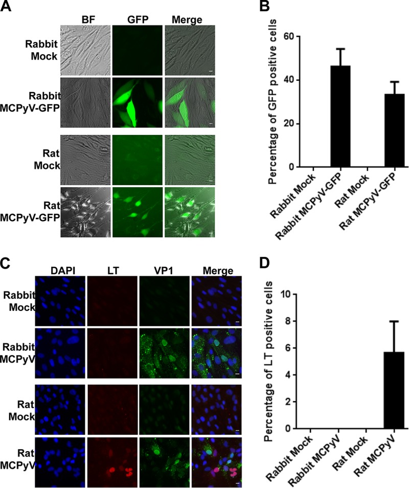
FIG 2.

MCPyV-GFP and MCPyV infection of dermal fibroblasts isolated from rabbit and rat dermal fibroblasts. (A) Dermal fibroblasts isolated from rabbit and rat skin samples were treated with MCPyV-GFP pseudovirions as described in the legend to Fig. 1A and examined under a fluorescence microscope. Bar, 10 μm. (B) Percentages of GFP+ cells in the experiment whose results are shown in panel A. (C) Dermal fibroblasts isolated from rabbit and rat skin samples were treated with MCPyV virions and stained as described for Fig. 1C. Bar, 10 μm. (D) Percentages of LT+ cells in the experiment whose results are shown in panel C. Error bars represent SEM of the results of at least three independent experiments.
