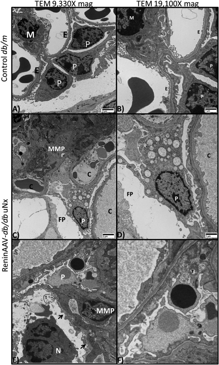
Figure 4.
ReninAAV db/db uNx mice exhibit ultrastructural changes observed in DKD. (A and B) Transmission electron micrograph of glomerulus from a healthy db/m control kidney with numerous intact podocytes (P) and basement membranes lined by intact endothelial cells (Es) with normal fenestrations. (A and B) A single mesangial cell (M) appears in the upper left quadrant. (C–F) Transmission electron micrograph of glomerulus from a ReninAAV db/db uNx mouse. (C and D) Extensive mesangial matrix proliferation (MMP) composed of amorphous and occasional fibrillary densities; three capillary profiles (Cs) are shown with thickened endothelium and irregular basement membranes with fused and expanded P foot processes (FPs). (C–F) Ps undergoing vacuolar degeneration; reactive endothelium lining capillaries (arrows) and neutrophil (N) infiltration were detected in the ReninAAV db/db uNx mice. n=6 mice evaluated per group. Scale bar, 1 or 2 μM.

