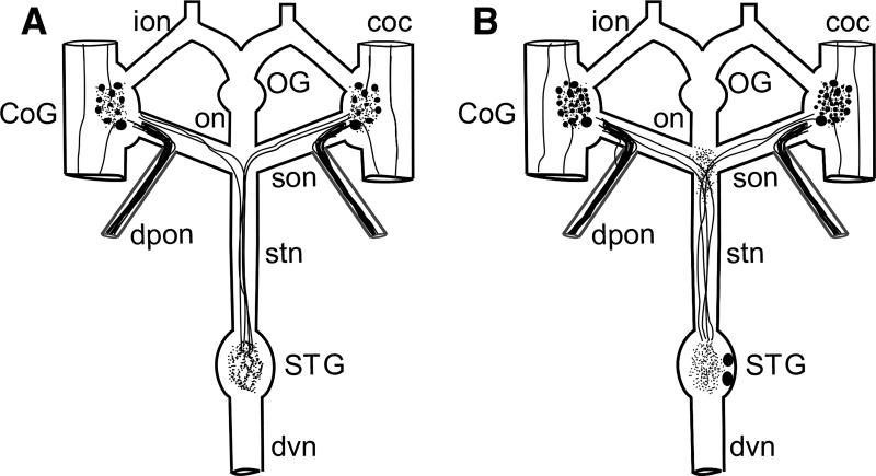
Fig. 2.
Schematic representation of the Homarus americanus stomatogastric nervous system (STNS) and the distributions of AST-C I/III and AST-C II in the lobster STNS. a Schematic representation of AST-C I/III-like immunoreactivity in the STNS. In this portion of the nervous system, the AST-C I/III antibody labels approximately 14 somata in each commissural ganglion (CoG), neuropil in each CoG and in the stomatogastric ganglion (STG), and axons in the circumesophageal connectives (cocs), superior esophageal (son), dorsal posterior esophageal (dpon) and stomatogastric (stn) nerves. No AST-C I/III-like labeling is present in the OG, nor is immunoreactivity present in the inferior esophageal (ion), esophageal (on) or dorsal ventricular (dvn) nerves. b Schematic representation of AST-C II-like immunoreactivity in the STNS. In the STNS, AST-C II-like labeling is present in approximately 42 somata in each CoG and two somata in the STG. Labeled neuropil is present in each CoG, in the STG, and at the son/stn junction. Labeled axons were seen in the cocs, sons, dpons, and stn. No AST-C II-like labeling was present in the OG, nor was any present in the ions or dvn. In both a and b, filled circles represent immunopositive somata, thick lines within nerves represent immunopositive axons, and tangles of thin lines represent regions of immunopositive neuropil

