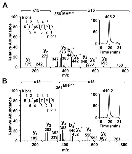
Fig. 1.
Identification and quantitation of MP20 168–173 pSer170. MS/MS spectra from an 18 year old lens confirmed the identity of A) endogenous MP20 168–173 pSer170 and B) its isotopically labeled AQUA peptide internal standard. Within the MS/MS spectra, the asterisks represent loss of phosphoric acid. The peptide sequence and fragmentation are shown in the top left corner of each panel, where phosphorylation is indicated by “p” and the isotopically labeled amino acid is indicated by “l” (bottom panel only). The XICs of MP20 168–173 pSer170 (panel A, insert) and its AQUA internal standard (panel B, insert) show the difference in mass between the (M+2H)2+ ions for the endogenous and labeled peptides and the identical elution times of these peptides.

