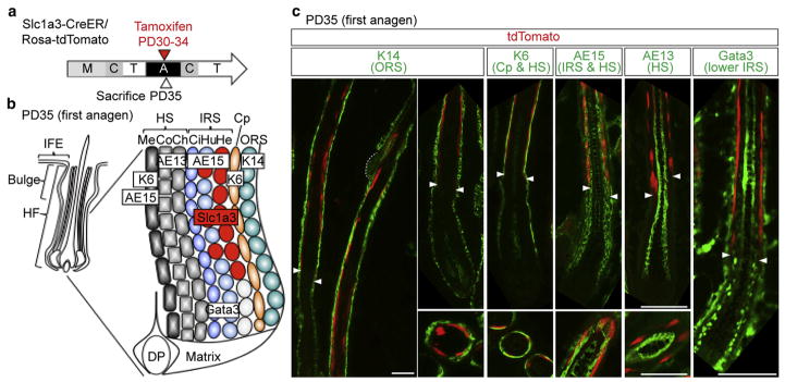
Figure 1. Slc1a3-CreER specifically marks the inner root sheath in the anagen follicle.
(a) Scheme for lineage tracing of Slc1a3-CreER marked cells. (b) Structure of the anagen hair follicle. Appropriate markers for each layer are shown in boxed text. (c) Immunostaining with lineage markers indicated in (b). Upper panels show sagittal sections and lower panels show cross sections of the hair follicle. The dashed line indicates the bulge.
Arrowheads represent the boundary between the upper and lower IRS. Scale bars = 50 μm. A, anagen; C, catagen; Ch, cuticle of hair shaft; Ci, cuticle of IRS; Co, cortex of hair shaft; Cp, companion layer; DP, dermal papilla; He, Henle’s layer; HF, hair follicle; HS, hair shaft; Hu, Huxley’s layer; IFE, inter-follicular epidermis; IRS, inner root sheath; M, morphogenesis; Me, medulla; ORS, outer root sheath; PD, postnatal day; T, telogen.

